Ultrasound is one of the most widely used imaging exams in medicine, thanks to its safety, speed, and reliability. It is based on high-frequency sound waves that allow real-time visualization of internal organs, soft tissues, and blood vessels. Without using radiation, it is a first-line diagnostic method to assess different areas of the body, monitor diseases, and support medical procedures.
What is an Ultrasound and what is it for?
Ultrasound uses high-frequency sound waves emitted by a probe and transmitted through tissues, which reflect them to form an image of the examined area.
At X-Clinic, ultrasounds are performed with the latest technology, using High-Resolution Digital Ultrasound equipment, including three-dimensional technology and real-time 3D visualization (4D), Color Doppler, and panoramic imaging.
It is a simple, safe, non-invasive, and painless technique. It does not use ionizing radiation and is therefore considered a harmless diagnostic method, including for children and pregnant women.
Ultrasound is used in different areas of medicine to:
- Assess the structure and function of organs and tissues;
- Support early diagnosis of diseases;
- Monitor treatments or surgeries;
- Carry out screening and preventive follow-up exams.
How much do ultrasounds cost?
The price of most ultrasounds performed at X-Clinic on a private basis is between €50 and €60 (out-of-pocket). However, X-Clinic has the following agreements:
- ADSE (the patient only pays the co-payment of €6 a €8), ADM GNR, SAD PSP;
- Insurance companies: Médis, Multicare, Advance Care, Allianz, Saúde Prime, Future HealthCare, SS CGD, SAMS Quadros, SAMS, Montepio, RNA, Generali, Mudum, MGEN, Aegon, Vitória, Una Seguros, other insurers;
- Health Plans: Medicare, Saúde Prime, Future HealthCare, Continente Wells, ACP, other health plans.
The final price varies depending on the healthcare system or insurance you want to use.
How is it performed?
During an ultrasound, the patient lies on an examination table in a dimly lit room to facilitate image reading.
The doctor applies a transparent gel to the skin, which enables proper transmission of sound waves and allows the probe to glide smoothly.
Moving images are observed in real time on a screen and interpreted by a doctor specialized in medical imaging.
In some cases, the probe may be inserted inside the body (endovaginal or transrectal), allowing a closer view of certain organs.
The procedure generally lasts between 10 and 30 minutes, depending on the type of ultrasound.
What does it detect?
In general, ultrasound is a very versatile exam that can detect structural and functional changes in different organs and tissues. It can identify masses, cysts, and nodules, as well as signs of inflammation or infection. With Doppler, it can also evaluate blood flow, which is useful in the study of vascular diseases. This exam also enables the detection of stones and obstructions in organs such as the kidneys or gallbladder, as well as changes in the morphology of various internal organs. Depending on the area assessed, ultrasound helps determine the presence, location, extent, and characteristics of lesions, and it is also a key tool for monitoring the evolution of conditions over time.
Types of Ultrasound Scans
Ultrasound is a versatile exam, indicated in many clinical situations. At X-Clinic, you can undergo several types of ultrasound, namely:
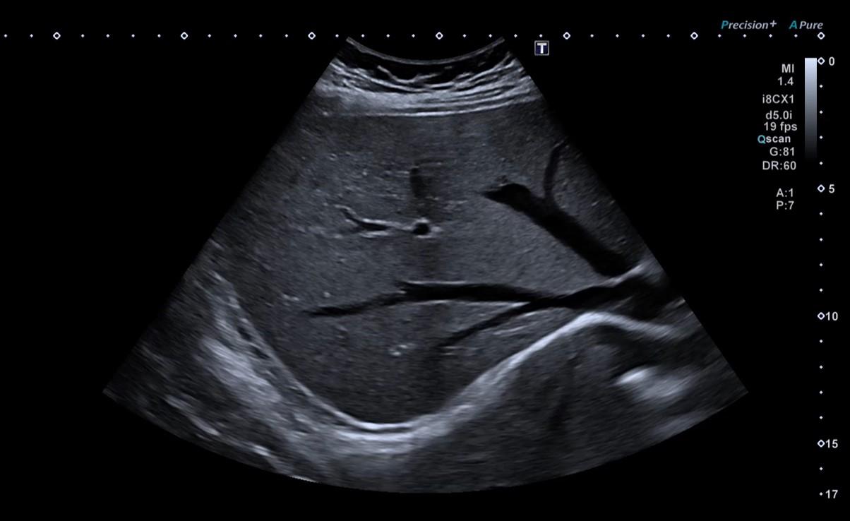
- Abdominal ultrasound: evaluation of the liver, gallbladder, pancreas, spleen, and other abdominal organs.
- Scrotal/testicular ultrasound: evaluation of the testicles, epididymis, and scrotal sacs.
- Breast ultrasound: study of the mammary glands and differentiation of solid or fluid-filled nodules.
- Pelvic/gynecological ultrasound (suprapubic and endovaginal): evaluation of the uterus, ovaries, fallopian tubes, and pelvic vessels. There are two techniques to observe pelvic organs: the suprapubic technique (the probe is placed on the skin of the lower abdomen) and the transvaginal or endovaginal technique (the probe is placed inside the vagina).
- Prostate ultrasound (suprapubic and transrectal): evaluation of the shape, texture, and integrity of the prostate. Transrectal prostate ultrasound, performed by inserting a small probe into the patient’s rectum, is an excellent exam for prostate assessment and for diagnosing prostate carcinoma.
- Renal and adrenal ultrasound: evaluation of the kidneys regarding their morphology and density, as well as the urinary tract. It also allows detection of possible nodules or masses, kidney stones, or urinary obstructions.
- Thyroid ultrasound: evaluation of thyroid volume, texture, and nodules.
- Suprapubic bladder ultrasound: visualization of the bladder, prostate, and seminal vesicles, performed via the suprapubic route.
- Salivary gland ultrasound: detection of nodules, cysts, or stones in the parotid, submandibular, and sublingual glands.
- Musculoskeletal (or joint) ultrasound: study of various joint structures, such as the shoulder, knee, elbow, and ankle, as well as muscles, tendons, ligaments, bursae, synovial membranes, and menisci.
Preparing for an Ultrasound
Preparation varies according to the area being studied:
- Abdominal ultrasound: fast for 6 hours before the exam; you may take your usual medication.
- Scrotal/testicular ultrasound: no prior preparation.
- Breast ultrasound: no prior preparation.
- Pelvic/gynecological ultrasound (suprapubic and endovaginal): full bladder; do not urinate 1 hour before, or drink 1 liter of water one hour prior to the exam.
- Prostate ultrasound: 1) Suprapubic: drink 1 liter of water one hour before; 2) Transrectal: perform 2 cleansing enemas one hour before leaving home.
- Renal and adrenal ultrasound: no prior preparation.
- Thyroid ultrasound: no prior preparation.
- Suprapubic bladder ultrasound: drink 1 liter of water before the exam.
- Salivary gland ultrasound: no prior preparation.
- Musculoskeletal ultrasound: no prior preparation.
In all cases, bring previous exams (radiological and laboratory), whenever available, and make sure to bring the exam prescription.
Where to have it done?
Ultrasound can be performed at X-Clinic (Av. Eng. Duarte Pacheco, nº26 – Mezzanine floor, 1070-110 Lisbon) or at NRD (Avenida Columbano Bordalo Pinheiro, nº 11-B, ground floor, 1070-060 Lisbon – next to Praça de Espanha).
